Deniz Publication
Clinical Cancer Investigation Journal
ISSN Print: 2278-1668, Online: 2278-0513
ISSN Print: 2278-1668, Online: 2278-0513
Present study is an attempt to provide an alternative trial of the traditional therapeutic protocols used for cancer treatment, which result in many serious side effects that may lead to death. Plasma medicine is a multidisciplinary field of research combining biology and clinical medicine with plasma physics and chemistry to launch a new cancer treatment modality. Stevia extracts, a natural sweetener derived from the Stevia plant, have anti-cancer properties. Therefore, the current study investigates the outcome of Gliding Arc Discharge plasma (GAD) and Stevia's effects on the HepG2 cell line. Eight groups were categorized as the control group, Stevia extract group (17.5μg/ml), GAD exposed groups with three dose intervals (40, 60, and 80 sec) and combined groups, Stevia extract with each GAD dose. Cell viability, P53 and Bcl2 genes, PARP-1, and TNF-α protein expression were investigated. Results indicated that the most effective treatments occurred in the combined group of GAD exposure with 60 and 80 sec. that record significantly decrease in cell viability of the HepG2 cell line and enhancement of P53 up-regulation, Bcl2 down-regulation, and inhibition of both PARP-1 and TNF-α protein expression. In Conclusion, the current study showed that GAD exposure and Stevia treatment could achieve enhanced tool therapy for some types of cancer through molecular mechanisms of action. Cell line toxicity, activation of the tumor suppressor gene (P53), apoptotic regulator gene (Bcl2), DNA repair gene as PARP-1, and immune editing of TNF-α gene are the main effects of GAD combined with Stevia.
Within the broad category of disorders known as cancer, aberrant cells can begin to grow out of control, invade neighboring regions of the body, or spread to other organs from the point of origin in practically any organ or tissue in the body. The latter process, known as metastasizing, is a primary contributor to cancer-related deaths.[1]
Hepatocellular carcinoma (HCC), which is one of the most common liver cancers, accounts for 75–85% of liver cancer cases and is the most frequent kind of the disease. Liver cancer ranked sixth in incidence (841,000 cases) and fourth in fatalities (782,000 cases) worldwide in 2020.[2]
Cancer treatment has always been a challenging process. There have been uses of traditional treatment modalities such as radiotherapy, chemotherapy, and surgery. At the same time, there have been significant developments recently, which are nanoparticles, natural antioxidants, ablation therapy, stem cell therapy, targeted therapy, chemo-dynamic therapy, radionics, ferroptosis-based therapy, andsono-dynamic therapy.[3]
GAD therapy is a promising approach for treating cancer, which can selectively kill cancer cells while maintaining healthy cells. Reactive oxygen and nitrogen species (RNS and ROS) produced during plasma treatment are the leading causes of cancer cells being killed. In addition to its
ability to target cancer cells specifically, GAD treatment has been demonstrated to have anti-angiogenic, anti-inflammatory, and immune-modulatory capabilities, which may further boost its therapeutic potential. The effectiveness of this therapy in combination with other cancer therapies like immunotherapy, radiation therapy, and chemotherapy has also been shown to improve the overall effectiveness of cancer treatment.[6] Therefore, the present study combines GAD treatment with a natural effective product such as plant extract Stevia. Stevia is a plant extract, that has been studied on various cancer cell types and may be an effective anti-cancer agent. The plant extract contains carotenoids and polyphenols that affect normal cell growth, proliferation, and differentiation as well as cancer-related epigenetic dysfunctions, preventing metastasis, decreasing tumorigenesis, and/or enhancing the effectiveness of chemotherapy and radiotherapy.[7]
Stevia Bertoni is known for having potent antioxidant, antibacterial, anti-diabetic (antihyperglycemic, insulinotropic, and glucagonostatic), antiplatelet, anti-cariogenic, and anticancer properties.[8]
It was reported that the P53 transcription factor is a significant regulator of various cellular functions. P53 is triggered in the presence of genotoxic stress to aid in DNA repair, cell cycle arrest, and apoptosis.[9] One of the genes with the highest frequency of mutations in liver cancer is the tumor suppressor P53. To prevent carcinogen-induced tumor formation, P53 controls the expression of genes related to cell cycle progression, cell death, and cellular metabolism.[10]
Bcl2 protein is crucial in regulating apoptotic cell death. Apoptosis is frequently inhibited in various cancers due to excessive over-expression of pro-survival Bcl2 family members or abnormal suppression of pro-apoptotic Bcl2 family proteins. Furthermore, the discovery of cancer therapeutic drugs finds the pro-apoptotic and pro-survival Bcl2 family proteins are appealing targets due to their crucial function in regulating apoptosis.[11]
In addition to several repair factors, there is an overview of poly (ADP) ribose polymerase (PARP), a protein implicated in DNA repair pathways, and its expression levels in liver cancer. It is one possible target. Treatment outcomes for patients may be enhanced by inhibiting this protein, which could also increase the effectiveness of existing therapies.[12]
Tumor necrosis factor-alpha (TNF-α) is a viable target for HCC treatment approaches. Treatments against TNF-α impeded the growth of HCC tumors by causing cell death and lowering pro-inflammatory cytokine levels.[13] TNF-α inhibits cancer cell proliferation and is released by cytotoxic macrophages recruited by plasma-induced reactive species.[14]
Cell Line (HepG2) is a human liver cancer cell line, obtained from the Genetic Engineering unit, National Research Center of Egypt.
The cell line was cultured in EMEM medium (ATCC) with the addition of 10% of Fetal Bovin serum (FBS, Thermo Fisher Scientific) as announced by the supplier. The culture was retained in a humidified 5% CO2 .The medium was changed once a week at Atmosphere 37C0
 .
.
Stevia plant extract (17.5 μg/ml) was purchased from Al Memaar Home Company for Agriculture Development and Agricultural Investment.
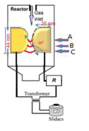
A GAD experiment design consisting of two identical diverging electrodes made of copper each with thickness (15mm), width (26mm), length (44mm), gap between two electrodes (1 mm), and arc (deviation of electrode) angle (120º). The electrodes are connected to an AC power supply (6.6kV) via a resistor (R=200kΩ) to avoid the high current. The input voltage was controlled with variable autotransformers (Slidacs).[15]
The AC power supply is a step-up transformer with a ratio of 1:30. The gas was injected from a narrow tube located ina narrow gap between two electrodes (Figure 1).
The gases utilized for the experiment were N2 and Ar where the gas flow rate was controlled using the pressure regulator. Discharge voltage was controlled using the voltage slide regulator and increased using the high voltage transformer (1:30), and then high voltage was utilized to the electrodes. Thus, the electric discharge frequency is 50 Hz. Discharge current was determined by both a Rogowski coil and clamp digital meter (for digital value) to connect to an oscilloscope (for following the current oscillogram).
|
|
|
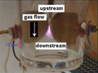
A: Gas Breakdown region B: Quasi-equilibrum (thermal) plasma region C: Non-equilibrum (non thermal) plasma region |
|
a) |
|
|
|
b) |
|
Figure 1. a) Gliding arc schematic and the electric scheme, b) GAD plasma photo. |
The samples were divided into eight groups and the treatment was as follows: First Group: Control sample group that is non-treated (HepG2) cell line (G1).Second Group: The cell line was treated with Stevia plant extract with a concentration of (17.5μg/ml)[16] (G2).Third Group: The cell line was exposed to 40 sec. of GAD (G3).Fourth Group: The cell line was exposed to 60 sec. of GAD (G4).Fifth Group: The cell line was exposed to 80 sec. of GAD (G5).Sixth Group: The cell line was treated with Stevia extract and then exposed to GAD with a dose of 40 sec. (G6).Seventh Group: The cell line was treated with Stevia extract and then exposed to GAD with a dose of 60 sec. (G7).Eighth Group: The cell line was treated with Stevia extract and then exposed to GAD with a dose of 80 sec. (G8).
After treatment, the cell line was cultured for three days, followed by an MTT assay to determine the viability of the cell line.
Cell viability (%) = Mean OD/Control OD × 100.[17]
Molecular evaluations
To evaluate the impact of Extract and Plasma on the HepG2 cell line, a set of genes (P53 and Bcl2) and their primer sets are shown in Table 1. The treated and non-treated carcinoma cell line was collected. Afterward, RNA extraction and RT-qPCR were carried out to measure the expression levels of the genes of interest.
Total RNA from control and treated cells were extracted using the RNeasy Mini Kit (Qiagen) according to the manufacturer’s instructions.
RT-qPCR was performed using the SYBR Green PCR Master Mix (Fermentas, USA). Data are expressed as the expression relative to GAPDH mRNA as a housekeeping gene using the 2^-∆∆CT method.[18]
Table 1. Specific primer sequences used in RT-PCR |
||
|
Primer |
Direction |
Sequences 5’-3’ |
|
P53 |
F |
TAACAGTTCCTGCATGGGCGGC |
|
R |
AGGACAGGCACAAACACGCACC |
|
|
Bcl2 |
F |
TTGTGGCCTTCTTTGAGTTCGGTG |
|
R |
GGTGCCGGTTCAGGTACTCAGTCA |
|
|
GAPDH |
F |
GTCTCCTCTGACTTCAACAGCG |
|
R |
ACCACCCTGTTGCTGTAGCCAA |
|
The purpose of this procedure is to provide a set of initial circumstances for the Western blot analysis of cell lysate samples.[19-22]
The proteomics analysis procedure for the following genes which are the PARP-1 gene and the TNF-α gene as follows
Proteins that had been electrophoresed on SDS-PAGE were moved to a HybondTM nylon membrane (GE Healthcare) using a TE62 Standard Transfer Tank with a cooling chamber (Hoefer Inc.) and they were then allowed to incubate in blocking solution for one hour at room temperature. β-actin was also utilized as a housekeeping protein.
 . Anti- β-actin primary antibody (Abcam, ab8227) was utilized to normalize the results.
The data analysis process used the Gel documentation system (Geldocit, UVP, England) and total lab analysis software (www.tatallab.com, ver. 1.0.1).[23]
Data collected were subjected to statistical analysis through the CoStat software program to detect the significance between the samples. CoStat version 6.400, copyright © 2022-2008 COHORT SOFTWARE (798 Lighthouse Ave. PMB 320, Monterey, CA, 93940, USA).[24]
Results and Discussion
The current data showed the effects of the chosen treatment on cell viability Table 2. There is a significant decrease in cell viability percentages after all treated groups with Stevia and/or Gliding arc plasma exposure (G2, G3, G4, G5, G6, G7, and G8) when compared with G1. It was well noticed that the most significant value was recorded in G7 (combined treatment of stevia and 60 Sec plasma exposure) with a percentage of 26%.
The percentage values of all groups when compared with G1 as follows (G2, G6, G8, G4, G5, and G3) with percentages (34, 42, 43, 61, 63, and 74%) respectively.
Table 2. Cell viability % in HepG2 cell line treated with Stevia and/or Gliding Arc Plasma Radiation (40, 60, 80 Sec) |
|
|
Groups |
Cell Viability (%) |
|
Group (1): Control. |
90±3a |
|
Group (2): Stevia extract. |
34±1f |
|
Group (3): Plasma exposure at 40 Sec. |
74±3b |
|
Group (4): Plasma exposure at 60 Sec. |
61±2d |
|
Group (5): Plasma exposure at 80 Sec. |
63±0c |
|
Group (6): Combination of Stevia extract and plasma exposure at 40 Sec. |
42±2e |
|
Group (7): Combination of Stevia extract and plasma exposure at 60 Sec. |
26±1g |
|
Group (8): Combination of Stevia extract and plasma exposure at 80 Sec. |
43±1e |
Significance level variance increased from (a) to (g) when compared with G1 (Costat statistical analysis).
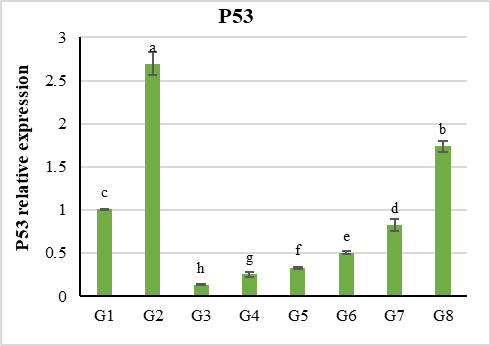
Figure 2 displayed the result values of relative gene P53 expression recorded in descending order from a to h when compared with each other according to the statistical analysis. The most significant expression values of the P53 gene were detected in G2 and G8.
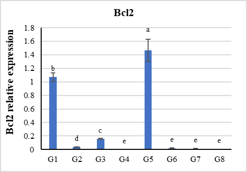
Whereas Bcl2 gene expression displayed a descending order from (a) to (e) when compared with each other according to the statistical relative analysis. All groups recorded significant depressed values of expression for the Bcl2 gene except the value in G5.
|
|
|
a) |
|
|
|
b) |
|
Figure 2. Levels of P53 and Bcl2 genes relative expressions in HepG2 cell line treated with Stevia and/or Gliding Arc Plasma radiation (40, 60, 80 Sec) |
PARP-1 is one of the main epigenetic proteins in the processes of transcription, and repair of gene expression. Tumor Necrosis Factor-α (TNF-α) represents the primary cytokine for tumor pathways. Their roles via effects of Stevia or gliding arc plasma exposure were measured by Western Immunoblotting techniques which were described by computerized Dendrogram for the gel analysis.
The lane keys of the groups for PARP-1 and TNF-α
|
Lane |
Lane 1 |
Lane 2 |
Lane 3 |
Lane 4 |
Lane 5 |
|
Groups |
Control |
40 second |
60 second |
80 second |
Stevia Extract |
Figure 3 characterized the bands of all groups for PARP-1 protein expression revealing that the most expression was recorded at lanes 4 and 5, while the least expression occurred at lanes 2 and 3 when compared with control lane 1.
|
5 |
4 |
3 |
2 |
1 |
|
|
||||
|
Figure 3. represented the bands of all groups for PARP-1 protein expression |
||||
By using the Dendrogram of PARP-1 protein expression its level values are represented as follows78.65, 64.70, 70.65, 90.44, and 99.25 for lanes 1, 2, 3, 4, and 5, respectively (Table 3).
Table 3. Data Parameter ofPARP-1protein expression level for HepG2 cell line protein with the four treatments |
||||
|
Lane 1 (%) |
Lane 2 (%) |
Lane 3 (%) |
Lane 4 (%) |
Lane 5 (%) |
|
78.65 |
64.70 |
70.65 |
90.44 |
99.25 |
Figure 4 represented the bands of all groups for TNF-α protein expression revealing that the most pronounced effect on expression was recorded at lane 2 and the least expression occurred at lane 5 when compared with control lane 1.
|
5 |
4 |
3 |
2 |
1 |
|
|
||||
|
Figure 4. TNF-α protein expression level for HepG2 cell line protein with four treatments. |
||||
By using the Dendrogram of TNF-α protein expression its level values are represented in Table 4 as follows 79.91, 81.91, 78.04, 75.21, and 67.75 for lanes 1, 2, 3, 4, and 5, respectively.
Table 4. Data parameters for level of TNF-α protein expression of HepG2 cell line protein for the fourth treatment. |
||||
|
Lane 1 (%) |
Lane 2 (%) |
Lane 3 (%) |
Lane 4 (%) |
Lane 5 (%) |
|
79.91 |
81.91 |
78.04 |
75.21 |
67.75 |
The bands of all groups for β-actin protein expression that was used as a reference and its values were 100% for all lanes.
Recently, there has been another voice depending on several confounding factors to verify cancer definition. To define cancer, we must take many considerations and all factors surrounding the cells as checkpoints, chromosomal instabilities, chemical factors, physical factors, psychological stress, failure of immunological recognition, etc. This leads us to postulate that it is essential to take into consideration all the aforementioned factors in dealing with the concept of cancer and its treatment or even its assessment. The most direct and effective promoter for all these factors is the epigenetic factor especially the DNA's physical nature that regulates its gene expression without changing its sequences.[25]
Cell lines are in vitro model systems that are often employed in many medical research domains, particularly drug discovery and basic cancer research. Their capacity to offer a never-ending supply of biological material for research is what makes them so valuable. When maintained in a suitable environment and with the necessary controls, verified cancer cell lines maintain the majority of the genetic characteristics of the original malignancy.[26]
Plasma radiation represented an effective option for the treatment of heat-sensitive materials and animal or human tissues.[27] Beneficial effects have been investigated in medical conditions variety such as breast, ovarian, pancreatic, melanoma, glioblastoma, osteosarcoma, and cervical cancer, etc. It is assumed that the main mechanism of the plasma’s effect is mediated using a stimulating dissipation of energy via chemical and/or radiation energy. It has been reported for various sources and cell lines that plasma can affect nucleic acids. In addition, the exposure to plasma in vivo did not show adverse effects in animals or small clinical cohorts in humans.[25]
Moreover, in preclinical or clinical settings, a variety of natural substances are employed to treat cancer. Then combining these natural products with GAD plasma treatment may augment its efficacy. The majority of natural medicine usually impacts many pathways, such as initiating apoptosis or preventing cell division, among other effects. Natural products can target several targets, which enables them to effectively counteract the biological complications associated with cancer and provide advantageous resources for cancer chemoprevention.[28]
Stevia is characterized by unique and effective ingredients as different glycosides, flavonoids, and fatty acids are among them, and they work together to give the plant its wide range of biological benefits. These ingredients permit Stevia products to have chemotherapeutic effects on cancer, treat polycystic kidney disease, increase insulin production in diabetics, and have potent antibacterial, antioxidant, and immunomodulating properties.[29]
The present study assesses the cell viability and the anti-cancer and anti-inflammatory effects of GAD plasma irradiation and/or Stevia extract in hepatocellular carcinoma cell lines (HepG2).
Table 2 showed a significant decrease in HepG2 cell line viability percentages after all treated groups with Stevia and/or Gliding arc plasma exposure (G2, G3, G4, G5, G6, G7, and G8) when compared with G1.
The most effective decrease in cell viability percentages was recorded in all combined groups (G6, G7, and G8). While G7 which represents treatments of Stevia and 60 Sec plasma exposure evidenced the least value for cell viability (26%). Two types of genes that have an important role in inducing the death and proliferation of malignant cells were selected in this study, which are P53 and Bcl2 genes. The P53 gene is one of the tumor suppressor gene classes. That has an important and basic role in the cell-programmed death operation and the death of cells by apoptosis. The P53 protein is a transcription agent that can trigger the expression of multiple target genes and plays vital roles in regulating genomic stability, apoptosis, and cell cycle, and is widely considered the “guardian of the genome”.[30]
Apoptosis, a programmed cell death pathway, is a critical physiological mechanism that guarantees cellular homeostasis and entirely cellular well-being. In the cancer context, where apoptosis evasion is a hallmark, the over-expression of anti-apoptotic proteins like Mcl-1, Bcl-xL, and Bcl2, has been documented. Accordingly, these proteins have transpired as promising targets for therapeutic interventions.[31]
In this context, Figure 2 displayed the results of relative values for P53 and Bcl2 gene expression. The most significant expression values of the P53 gene were detected in G2 (Stevia alone) and G8 (Stevia and Plasma with dose 80 sec.). Whereas, Bcl2 gene expressions displayed significantly decreased values with all treatments when compared with the control group except the value of G5 (Plasma dose 80 Sec.).
The variation in the level of malignant cell death (HepG2 cell line) and on the level of the two genes transcription may be returned to that the two treatments work by different mechanisms, where Stevia extract works chemically. In contrast, Plasma works physically and its radical constitutes effects.
A class of similar enzymes known as poly ADP-ribose polymerases (PARPs) can catalyze the delivery of ADP-ribose to target proteins. DNA repair, recombination, replication, and transcription, chromatin structure are just a few of the biological functions that PARPs are crucial to. Additionally, PARP inhibitors may make tumors more vulnerable to chemicals that damage DNA.[32]
In recent years interest in studying drugs targeting mutations in various mechanisms involved in DNA repair is increasing: Genes and PARP associated with DNA homologous recombination repair are crucially involved in the protecting cells process from DNA-damaging agents such as ionizing radiation or chemotherapy.[33]
In this framework, the current study was designed to determine which of the two treatments separately affects the inhibition of repair processes in cancer cells, Stevia or plasma radiation. Obtained results in Table 3 declared that Plasma radiation is more effective than Stevia treatment for inhibition of PARP especially for doses 40 and 60 sec. These findings were discussed and interpreted given the Plasma radiation's unique effect as an epigenetic agent and its role in controlling the transcription process.[25]
Given these facts, the TNF-α effect depends on cell types (normal and cancerous cells) and the consequences of its promoter signals. Then in the present study protein, expression data of TNF-α was measured in HPG2 cancer cells that induce growth and proliferation. The most effective treatment is Stevia, Plasma radiation with 80 and 60 Sec. Table 4 respectively. This finding may be attributed to the ability of Stevia and Plasma doses (80 and 60 Sec.) to induce pathways to inhibit TNF-α overproduction via its miscellaneous signaling pathway.[34]
According to the gained data, gliding arc plasma and Stevia may be considered promising tools for cancer treatment (particularly combined treatments with Stevia and high Plasma radiation doses) via varieties of pathways and modes of actions as will be declared in the following discussion.
For Plasma, application to biological cellular structures has been reported to generate abundant reactive species. Their interaction with cells manipulates cellular redox signaling, finally resulting in alters in surface receptor function, cell cycle arrest initiation, P53activation using DNA damage, and subsequent P53-dependent apoptosis and other impacts on different cells.[35, 36]
There are several types of Plasma according to their applications. Only plasma produced at moderate temperatures and atmospheric pressures should be used in plasma-assisted medicine. GAD uses an electric field as an excitation source. Because of the reactor type and the discharge utilized, there are many GAD generation sources, including DBD (dielectric barrier discharge), glow, and gliding or corona discharge. Regardless of the GAD generation method, its main benefits are low cost, the process controllability, and the easy switch–on–switch–off ability.[37]
It is a global challenge to find a precise cancer treatment because there are plenty of cancers and each one needs a different course of action. A common treatment that is effective despite having numerous negative side effects is chemotherapy. Certain cancers even develop resistance to this treatment. To mitigate these negative side effects, researchers are also creating novel, potent therapies. Science has found a new cancer treatment which is GAD that works similarly to radiation and chemotherapy. GAD produces ions, electrons, radicals, and other new species through an ionization process that has anti-cancer properties and the ability to destroy malignant cells.[6]
Based on preceding similar works on different types of plasmas and their impacts on both normal and cancerous cells, their obtained data evidenced that atmospheric non-thermal plasma and gliding arc plasma exposure can be utilized for repair tissues, cure some types of diseases and treat tumors especially cases that have drug-resistant.[38, 39]
As well, they stated that the most effective role of plasma was the significant increase in the chromosome instabilities arrested using induction of various mitotic checkpoints ended by necrosis and apoptosis processes. Additionally, the same events were detected for the transcriptional levels of anti-tumor genes (Bcl2, caspase-3, and P53genes) and enhancement of immunological responses (TNF-α and interleukins).[40] Moreover, plasma affects the local microenvironment of cancerous cells, especially in the extracellular matrix by stopping cell spreading, lyses, and eradicating cell-to-cell communications.[41]
Other obstacles still facing cancer treatment strategies: cancer cell defense therapy is killing normal cells. The immune system acts closely with tumor cells in the host, contradictory promoting and inhibiting tumor progression and development. There are three stages to this process, which is called cancer immunoediting: elimination, equilibrium, and escape. GAD aids and promotes this process of immunoediting of cancerous cells.[42]
Overview of the obtained results impresses that HepG2 cell line exposure to GAD arrested the repair system and restricted the spreading of cancerous cells (metastasis) through up and down-regulation of protein expression for some types of cytokines and immunological mediators, especially at 60 and 80 Sec time exposure. It was confirmed that TNF-α is a main mediator of cancer-related inflammation. So, treatment with TNF‑α antibody markedly decreased the levels of these pro-inflammatory cytokines due to that inflammation is closely associated with HCC. Consequently, anti-TNF-α treatment may have therapeutic value in HCC treatment by assisting to neutralize the pro-inflammatory microenvironment. Plasma irradiation reduces all these inflammation factors or mediators.[13]
In addition, recent studies can interpret plasma strategies for cancer treatment through both oxidative stress in cancer cells (free radicals) and epigenetic factors effects via its resonance nature of electrons.[25]
Other studies discussed the mechanism of action for the Stevia plant which contains a high percentage of rebaudioside and steviol glycoside (stevioside). Stevioside has a potent inducer for apoptosis and it conveys the apoptotic signal through intracellular ROS generation; there by inducing a pathway of mitochondrial-mediated apoptotic. Overall, our data revealed that stevioside induces the ROS-mediated mitochondrial permeability transition and results in the enhanced expression of apoptotic proteins such as Caspase-9, Bcl2, and Bax.[43]
In light of these aforementioned literature studies, our results declared that Plasma medical applications supported with some natural products such as plant extracts such as stevioside and the other Stevia constitute have strong anticancer action in cultured HepG2 cell lines. This occurred through a multiple-action combination, including DNA synthesis inhibition, cell viability suppression, and induction of apoptotic cell death via the mitochondrial apoptotic pathway.
None
None
None
None
|
||||||||