Deniz Publication
Clinical Cancer Investigation Journal
ISSN Print: 2278-1668, Online: 2278-0513
ISSN Print: 2278-1668, Online: 2278-0513
Breast cancer stands as a pervasive cause of mortality among women globally, necessitating comprehensive investigations into its complex development and progression. This study aims to comprehend the complexities of breast cancer development, focusing on the roles of cell multiplication, movement, and the impact of cisplatin as a therapeutic agent. Additionally, it explores the significance of interleukin-6 (IL-6) in breast cancer, emphasizing the analysis of IL-6 gene expression changes pre and post-treatment using MTT assays and reverse transcriptase techniques. The research reveals that cisplatin effectively inhibits breast cancer cell proliferation, showcasing its therapeutic potential. The analysis of IL-6 gene expression demonstrates significant changes, offering insights into the molecular alterations induced by cisplatin and its impact on key signaling pathways associated with cancer progression. Major conclusions include the promising role of cisplatin in impeding breast cancer cell proliferation and its influence on the molecular landscape. The reduction in IL-6 gene expression post-treatment underscores cisplatin's multifaceted effects. The study highlights IL-6 as a potential biomarker for treatment response prediction, contributing valuable insights for personalized breast cancer treatment approaches.
All over the world, breast cancer is what mainly kills women with cancer. Even though there are many ways to treat breast cancer like using radiation, special drugs targeted towards the disease, medicines that poison fast-growing cells, and surgery, a big part of these treatments fail. This often causes the spread or comeback of this type of cancer called metastasis. It is the most prevalent cancer among females. Its etiology is complex, its developmental trajectory can take decades, and its clinical course is wildly unpredictable.[1] Breast cancers have many types and are often sorted by the kind of cells they contain using tests like checking for prognostic markers such as HER2, PR, and ER.[2] These tell about how a patient's disease might progress. The amount and outcome of four types of breast cancer—Luminal A, B, HER2-enriched, or triple-negative —are different. Two tough types of breast cancer are HER2-enriched and triple-negative breast cancer (TNBC). They have worse results for the patient, and show how bad things might get by having a lot more Ki67 staining. It's harder to treat these cancers because they spread quickly to other parts of your body.[3] Most times when people die from this kind The most common places where breast cancer spreads are the liver, brain, lung, and bone. We use complex steps like changing cells to help them move around or multiply in a new part of our body for metastases.[4, 5] Breast cancer cells release cytokines and other small soluble proteins during many of these stages in an effort to prime microenvironmental cells and encourage cancer cells (autocrine effect) (paracrine effect). Proteins called cytokines are secreted and pleiotropic substances that regulate a wide range of inflammatory and immune reactions, many of which are exploited by cancer. IL-6 is one of several cytokines that can have both pro- and anti-cancer effects. IL-6 is a type of inflammation-triggering chemical made by different cells in the tumor area. This includes cancerous ones.[6] IL-6 is crucial for the growth and change of cancer cells. IL-6 levels in blood and tumor sites are higher than usual for some cancers like breast cancer. This is often linked to poor
We got MCF-7 breast cancer cells and normal breast cells from the National Centre for Cell Science (NCCS) in Pune, India. We then cultured them following the provided cell culture instructions. It was filled with 15% baby cow blood at a temperature of 37 ºC and had some CO2 air in a wet box room called an incubator.
To see how cisplatin affected breast cancer cells, they used the MTT test on the MCF-7 cell survivability. Shortly, cells called MCF-7 were put into a plate with 96 holes in the shape of circles. The number of them was one hundred thousand for each hole. After 12 hours, the cells were mixed with cisplatin for another 48 hours at a heat of 37 degrees. It was done while slowly raising the amount from 2.5 to full strong doses (from 0 to extremely high). We checked for the presence or absence of breast cancer cells. A normal medium was used as a control. All cells were grown for an extra 48 hours. Then, the MTT reagent was put into each little box and the cells were kept warm for four hours at 37°C. After the material was soaked and the liquid was taken off, 150 microliters of DMSO were put in to break up the dye. Then they checked how much light went through it at a number setting of "490 nm" on a tiny plate reading machine. The IC50 means the level of drug needed to cause 50% death in cells, compared with a normal control. In this trial, the IC50 was 20 micrograms per milliliter. The test was done three times; six similar samples were seen each time.[9]
The TRIzol reagent (Invitrogen, Carlsbad, USA) was used to get the total RNA from both cancerous and normal tissues. This is done as per what the maker's instructions say. The cleanness and amount of RNA taken out were checked using a Nanodrop 2000 Lite machine (Thermo Fisher Scientific in Waltham, MA). For more testing, the RNA was kept in a very cold place that is -20 degrees.[8, 10, 11]
A total of 10 liters was used within the Moloney Murine Leukemia Virus (M-MLV) reverse transcriptase. This process then changed all RNA into complementary DNA or cDNA after being heated up and immediately chilled down. The mix is then put in a PCR (Thermo Fisher MiniAmp plus thermal cycler) for 10 minutes at 30°C. After that, it spends 30 more minutes at the temperature of 42°C followed by an extra five long ones to get warm up fully on reaching and staying safe within range from its starting point till finalizing We measure the amount of cDNA using Nanodrop lite and store it at -20°C until we study more details. The gene TGF- was studied for how well it makes proteins from the cDNA using a dye called SYBR Green (Takara, Japan). They looked at GAPDH as a normal control. TGF- primer sequence (forward and reverse) and GAPDH primer sequence (forward and reverse). To amplify all of the samples in duplicate, the following thermocycling settings were used: Melting at 95 degrees for half a minute, then repeating it 40 times by using heat of this degree again to mix everything; staying there each time starts with faster churning and finishes in slower stirring. Lastly, TGF expression was measured using the 2-Cq method.[12]
We used the TRIZOL agent to get the total RNA. We made DNA from a single chain in a mix. This included 2 milligrams of RNA, a primer called oligo-dT and the Superscript II reverse transcription tool. Real-time study (qRT-PCR) was done on a machine called iCycler, separately for each one. Using tested primers and SYBR Premix Ex Taq II. Using a cycle number limit, they measured how much certain genes were expressed. The gene GAPDH was used to make sure things were balanced in the same mixes.[8, 10]
The facts were shown as average standard deviation (SD). The student used a t-test program to compare the amount of TGF in cancerous and normal close-by tissue. P 0.05 was decided to be important in the field of statistics.
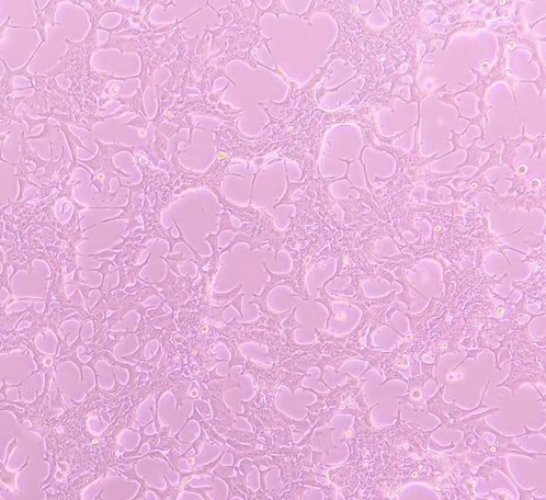
The microscopic analysis of breast cancer cells within the MCF-7 cell line reveals distinctive characteristics, notably the presence of keratin aggregates that serve as a hallmark feature associated with breast cell carcinoma. Through meticulous microscopic examination, the process of keratinization emerges as a pivotal factor contributing to the formation and growth of these breast cancer cells. This histological investigation (Figure 1) provides a nuanced understanding of the morphological aspects that define the pathological state within the MCF-7 cell line, offering valuable insights into the intricate features associated with breast cell carcinoma at the microscopic level.
|
|
|
Figure 1. represents breast cancer cells seen in the MCF-7 cell line. Keratin aggregates represent the formation of breast cell carcinoma. Keratinization causes the formation and growth of cells. |
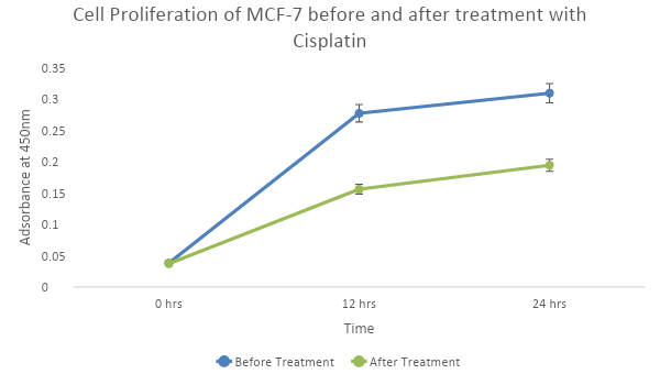
In investigating the functional impact of cisplatin on MCF-7 breast cancer cells, a detailed assessment of proliferation rates provides significant insights. The line chart (Figure 2) depicting cell proliferation dynamics before and after cisplatin treatment reveals a compelling narrative, unmistakably demonstrating a substantial reduction in the proliferative capacity of MCF-7 cells post-treatment. This finding underscores the potent inhibitory effect of cisplatin on the uncontrolled growth characteristic of breast cancer cells, presenting a promising avenue for therapeutic intervention. The observed modulation in proliferation dynamics highlights the potential of cisplatin to disrupt the aberrant cell division associated with breast cancer, emphasizing its role as a promising agent for targeted therapeutic strategies in breast cancer treatment.
|
|
|
Figure 2. illustrates the proliferation rates of cells before and after treatment with cisplatin. And as the graph shows the proliferation rate of MCF-7 cells has significantly reduced after treatment with cisplatin. |
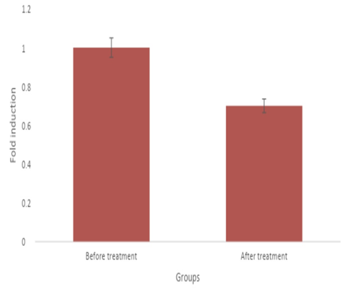
Delving deeper into the molecular realm, the gene expression analysis, highlighted in Figure 3, focuses on the IL-6 gene within MCF-7 breast cancer cells treated with cisplatin. The graph visually conveys a significant reduction in IL-6 expression following cisplatin treatment compared to normal breast cancer cells. This molecular modulation sheds light on the intricate influence of cisplatin on key signaling pathways associated with inflammation and cancer progression within MCF-7 cells. The observed decrease in IL-6 expression aligns with the broader therapeutic implications of cisplatin, suggesting not only a direct impact on cell proliferation but also a profound influence on the molecular landscape of breast cancer cells. This molecular insight strengthens the potential of cisplatin as a multifaceted therapeutic agent with implications for targeting molecular pathways crucial to breast cancer progression.
|
|
|
Figure 3. shows the expression of the IL-6 gene before and after the treatment of MCF-7 cells with cisplatin is seen. This graph shows that the expression of IL-6 was significantly reduced after the treatment with cisplatin when compared to the normal breast cancer cells. |
Cisplatin is thought to be one of the best cancer-fighting drugs that many people use for treating solid tumors. One of the best ways doctors use cisplatin is for treating breast cancer. Cisplatin is used to fight breast cancer through chemotherapy. Breast cancer is the main reason women around the world die. Surgery is also used to treat other types of cancers, but for breast cancer the only treatment choice beyond chemo and compulsory measures extensions for life is chemotherapy. Cisplatin-based chemicals used in chemotherapy have helped treat many types of breast cancer. This is because they attack fast-growing cells and cause harm as a side effect due to their toxic properties. But cisplatin makes it less harmful to cells. Most of its bad effects come from the creation and end of cell divisions by attaching cisplatin-DNA.[5]
In this study, breast cancer MCF-7 cells were given cisplatin, and said that it can greatly stop or reduce the growth of these cells. So, we know from studying this plant in labs how amazing the chemistry caveman times gave us is for making our lives full of good ways to get better nowadays! A study on 35 people shows that cisplatin is a powerful medicine for breast cancer treatment. It's also possible to pair it with other drugs in the first plans of care.[13, 14]
The main reason most women die from breast cancer is because it spreads to other parts of the body. This happens even though the initial tumor does not grow very big itself. IL-6 has been linked to the spreading of cancer in many lab studies. This link is made through different ways, including turning skin cells into other kinds and increasing how much a cell moves around or digs deep inside something. Also, it plays a role in attracting special types of healthy body immune system fighter cells called MSC recruitment which also comes from research settings! This IL-6 is a cytokine that takes part in the defense and swelling process. This increases when breast cancer cells are present.[15] In this study IL-6 gene expression in MCF-7 cells has been analyzed before and after treatment with cisplatin. And showed that the expression has significantly reduced after the treatment with cisplatin when compared to the normal breast cancer cells. The reduction in IL-6 gene expression after cisplatin treatment may be due to the drug and its effects on inflammation and immune responses. However, the specific effects on IL-6 gene expression may vary depending on factors such as the type of cancer being treated, the dose and duration of cisplatin therapy, individual patient responses, and interactions with other drugs or therapies.[16]
Several studies have shown IL-6 levels between the controls and breast cancer patients which can reveal the lower and higher stages of the disease. Another study checked IL-6 levels in breast cancer patients' blood serum to see if it linked with disease growth. They found out that breast cancer patients had more IL-6 in their blood compared to healthy women.[10]
Our study focused on elucidating the effects of cisplatin on the growth and migration of breast cancer cells, revealing a substantial reduction in both cellular proliferation and movement following treatment. Additionally, our investigation explored the molecular aspect by assessing the expression of the interleukin-6 (IL-6) gene, a key regulator of cancer growth. The observed decrease in IL-6 gene expression aligns with previous research, reinforcing cisplatin's efficacy in inhibiting breast cancer cell dynamics. These findings not only reaffirm the well-established impact of cisplatin on cellular behavior but also provide molecular insights into its ability to modulate critical pathways associated with cancer progression. Overall, our study underscores the multi-faceted therapeutic potential of cisplatin in mitigating both the proliferative and molecular aspects of breast cancer development.
Overall, the goal of future research is to deepen our understanding of basic mechanisms, optimize treatment strategies, identify biomarkers, and translate in vitro findings for clinical practice to improve breast outcomes for treated cancer patients’ cisplatin.
None.
None.
None.
None.
|
||||||||