Deniz Publication
Clinical Cancer Investigation Journal
ISSN Print: 2278-1668, Online: 2278-0513
ISSN Print: 2278-1668, Online: 2278-0513
Basal cell carcinomas (BCCs) are malignant, locally invasive, slow-growing tumors that arise from basal cells and represent 75% of non-melanoma skin cancers. BCC has many different clinical presentations, but the most common types include nodular, micronodular, superficial, morpheaform, infiltrative, and fibroepithelioma of Pinkus. These neoplasms develop in 80% of cases in sun-exposed areas of the head and neck, and only a minority of cases have been reported arising in the nipple-areola complex. In these locations, Paget’s disease of the breast has to be first ruled out before BCC could be considered. Furthermore, since most BCCs occur in adult age, cases that appear earlier in childhood with increased number and unusual distribution should be checked for genetic disorders such as Basal cell nevus syndrome.
Thus, we reported an original case of superficial BCC of the nipple-areola complex masquerading as Paget’s disease of the breast and revealing a Basal cell nevus syndrome.
Paget's disease of the breast (PDB) is a rare cancer of the nipple-areola complex (NAC) and is often found with underlying breast cancer.[1] It manifests as a burning or itching erythematous, crusted, or scaling lesion (eczema-like) and therefore can be mistaken for inflammatory skin conditions such as dermatitis and psoriasis as well as tumors like Bowen's disease, superficial spreading melanoma, and basal cell carcinoma (BCC).[2, 3] This latter is slow-growing and locally invasive skin cancer which derives from basal cells.[4] Although BCC is the most common human cancer worldwide, its occurrence in the NAC is exceptionally seldom.[5] Thus, it is widely admitted that DNA damage caused by ultraviolet exposure is the main pathogenesis factor involved in BCC carcinogenesis.[6] Therefore, while most BCCs develop on the sun-exposed skin of the head and neck areas, their appearance in the sun-protected regions, such as the breast, is rare and the NAC is further an extremely uncommon location. Approximately 74 cases have been reported in the literature till now.[7] Additionally, BCCs can occur either sporadically or in the context of hereditary génodermatoses such as Gorlin syndrome.[8]
Gorlin syndrome also known as Basal cell nevus syndrome (BCNS), is an autosomal dominant disorder caused by mutations in the PTCH1 (patched) gene with complete penetrance but variable phenotypic expressivity.[9] Finding commonly encountered in this disease, such as BCCs, odontogenic cysts, calcification of the falx, and ovarian fibromas, develop usually at different ages in affected individuals.[10] BCCs are the most frequent sign of BCNS.[11] The tumors are often multifocal and located primarily in sun-exposed areas but they are also increased in sun-protected ones. Relying mainly on clinical signs, diagnostic criteria proposed by Kimonis et al. help establish diagnosis and monitor disease progression.[8]
Here, we report a case of NAC cancer in an elderly woman, which was initially thought to be PDB, but later found to be BCC and revealed an undiagnosed BCNS.
A 68-year-old female patient was referred by her gynecologist to the dermatology department for a lesion on her right NAC of 6 months duration. She had no history of previous trauma, radiation exposure, or any potential risky behaviors. However, since her twenties, she has undergone multiple
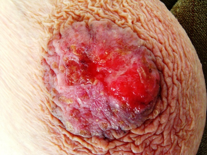
surgical excisions of BCCs, mostly located on her face and trunk. Physical examination revealed a 3 cm sharply defined, erythematous, annular, and firm plaque with ulceration on its surface that occupied the whole right NAC (Figure 1). There was no axillary lymph node involvement and the inspection and palpation of the left breast was unremarkable.
|
|
|
Figure 1. Basal cell carcinoma of the right nipple-areola complex showing ulceration. |
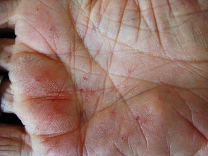
Further clinical examination found palmar pits (Figure 2), multiple milia on the face and fingers, and an epidermal cyst of the left thumb. The rest of the physical examination was normal, except for scars on her face and trunk resulting from previous surgeries. Thus, PDB was first considered, but there were no positive signs of mammary neoplasm detected on mammography as well and no axillary lymph node enlargement was noticed on the ultrasound exam.
|
|
|
Figure 2. Multiple palmar pits. |
The histopathological examination of a punch biopsy from the lesion consisted of multifocal, small nests of atypical basaloid cells, well-circumscribed and attached to the lower part of the epidermis. These nests remain confined, without apparent invasion, to the papillary dermis. Based on these histopathological findings, the diagnosis of superficial BCC was made.
Therefore, the lesion was excised. The combination of clinical symptoms and pathological findings fulfill the diagnostic criteria of BCNS namely two major criteria represented by multiple basal cell carcinoma recurring since her teenage years and more than three Palmar pits. No additional relevant findings of this syndrome were noted on physical and imaging examinations. The patient has been followed up for about three years and no clinical evidence of local recurrence or any other problem has been observed to date.
Results and Discussion
Non-melanoma skin cancers are the most common malignancies diagnosed in fair-skinned populations, with BCCs representing approximately 75% of all cases.[12] These neoplasms mostly develop on the sun-exposed areas of the head and neck, though, in rarer instances, they can arise in non-sun-damaged skin but exceptionally on the NAC. A literature review conducted by Chun et al. in 2016 [13] in the PubMed database, revealed 55 cases of BCC occurring in the NAC to this date. Since then about nineteen additional cases have been published. Based on these reported cases, we can draw two main characteristics of this particular location. Firstly, BCCs of the NAC are more frequently observed in males, likely because women are more unwilling to expose their entire chest to sunlight than men.[14] Of note, some authors have suggested that ultraviolet irradiation might be the main etiological factor, even for this uncommon location of BCC.[5] Secondly, they have more aggressive behavior compared to BCCs arising at other anatomical regions such as the head and the neck.[15] This poor prognosis is likely owing to the high rate of metastasis (4% - 3 / 74), though this statement was contested by some authors.[16]
BCC has many different clinical presentations, but the most common types include nodular, micronodular, superficial, morpheaform, infiltrative, and fibroepithelioma of Pinkus.[17] BCCs on the NAC present more frequently with nodular subtype (42.9%) whereas superficial ones, like our case, represent less than 31% of those seen in this region.[13] Clinically, a superficial BCC manifests as a red macule or thin plaque with indistinct margins and scaly or crusted surface and therefore can be confused with other neoplastic conditions such as Bowen disease or PDB but also with benign skin conditions such as dermatitis.[18] Indeed, due to clinical features, the initial diagnosis for our patient was PDB, but it has been subsequently ruled out by histological analysis.
On the other hand, although most cases of BCC are sporadic, in patients with BCCs occurring since childhood with increased number and unusual distribution, genetic disorders like BCNS should be considered.[3, 4]
This syndrome is a rare autosomal dominant multisystem disorder with complete penetrance and variable expression.[3] Although the spectrum of symptoms is broad, the main manifestations are BCCs, palmar or plantar pits, odontogenic keratocysts, and intracranial calcification. Therefore, specific diagnostic criteria for this syndrome have been developed, where it is necessary to meet 2 major criteria or 1 major criterion and 2 minor criteria to establish the diagnosis.[19] Thus, our patient had 2 major criteria including the presence of more than two BCCs with early onset and more than three palmoplantar pits.
We report a singular case of superficial BCC in many regards including the involvement of an uncommon location, its occurrence in the context of BCNS, and that clinically mimics PDB. Also, we would like to stress that no similar case of BCC arising on the NAC in the context of BCNS has not been reported in the literature before as far as we know.
I am grateful to the patient for allowing me to publish the case report and use her images. We would also like to acknowledge the collaboration of the Pathology and Radiology departments of the University Hospital Center of Constantine.
None.
None.
The study was approved by the Institutional Ethics Committee. Informed Consent for the publication of the case report and images was obtained from the patient. The patient’s identifying information has not been revealed in the report.
|
||||||||