Deniz Publication
Clinical Cancer Investigation Journal
ISSN Print: 2278-1668, Online: 2278-0513
ISSN Print: 2278-1668, Online: 2278-0513
Molecular Mechanisms Underlying Chemopreventive Anticancer Activity of Stevioside on Human Prostate Cancer Cell Line in vitro
Preethi Raj1, R Priyadharshini1*, Selvaraj Jayaraman2, Palati Sinduja1
1Department of Pathology, Saveetha Dental College and Hospitals, Saveetha Institute of Medical and Technical Sciences (SIMATS), Saveetha University, Chennai-600 077, Tamil Nadu, India. 2Department of Biochemistry, Saveetha Dental college and Hospitals, Saveetha Institute of Medical and Technical Sciences, Saveetha University, Chennai-600 077, Tamil Nadu, India.
Abstract
The burden of cancer incidence and mortality is rapidly increasing worldwide. The second most frequent cancer in men is prostate cancer which affects 1.41 million people worldwide (WHO statistics). Stevioside is an easily available item and it was observed to significantly inhibit cancer cell growth. Our study aims to analyze Molecular mechanisms underlying the chemopreventive anticancer activity of Stevioside on human prostate cancer cell lines. Prostate cancer cells were treated with Stevioside and the level of Bcl-2, Mcl-1, and Caspase-3 was estimated respectively. Data were expressed as the mean ± SD of 3 individual experiments performed in triplicate. Statistical analysis was performed using one-way ANOVA. In PC-3 cells, the effect of Stevioside extracts on cell viability was analyzed through MTT assay with a significant decrease in the percentage of viable cells with an increase in concentration, while the Mcl-1 gene with a decrease in fold change with a rise in concentration. Caspase 3 with a significant increase in fold change with an increase in concentration indicates effective apoptosis and also inhibits the Bcl-2 gene with a decrease in fold change with a rise in concentration with a significance of p<0.05. According to the results Stevioside extract considerably and strongly (by a significant fold change) suppresses the proliferation of cancer cells. The results imply that Stevioside may be turned into a natural prostate cancer medication and further investigation is necessary to establish the daily intake of Stevia products that is both safe and beneficial to the health of the human body.
Keywords: Anti-cancer, Natural, Prostate cancer, Stevioside, Good health, Well-being
Prostate cancer is a frequently diagnosed Invasive cancer. According to 2022 Prostate cancer statistics, hundred and twelve men out of one lakh per year new cases were reported of eighteen per one lakh men reported with an increased mortality rate.[1] Androgen deprivation is the current treatment protocol that has been followed which will generally respond for a shorter duration.[2] Apart from that, desirable control has been aided with chemotherapy. Apoptosis of tumor cells can inhibit the further progression of cancer and can be attained with a wide variety of natural and easily available products which can be attained as treatment drugs to inhibit further invasion and proliferation of cancer cells.[3]
Natural resources are a unique supply of diverse structural scaffolds that can yield effective chemical agents for the treatment of prostate cancer.[4] Stevioside is an easily available item and it was observed to significantly inhibit cancer cell growth. It boosts the body's urinary function and has hypotensive, vasodilating, sweetening, anti-fungal, anti-viral,[5] anti-inflammatory, anti-diabetic,[6] anti-bacterial, and anti-viral qualities.[7] It has been discovered that the plants in numerous nations, including Brazil, Japan, and Paraguay, are non-toxic, non-addictive, noncarcinogenic, and non-mutagenic. It has no teratogenic or genotoxic properties. It is safe for diabetics because it does not affect blood sugar levels.
However, the development of resistance to treatment and the danger of recurrence have prompted researchers to look for fresh anticancer agents, including those derived from therapeutic plants. Stevia rebaudiana, a plant species native to Paraguay and Brazil that is also used medicinally, produces stevioside in its leaves.[8, 9] About 70 of the many Stevia species are endemic to Mexico, and little research has been done on their anticancer properties. Their constituents, including flavonoids, sterols, and sesquiterpenes, which have significant biological activity, are responsible for these effects.[10] This study aims to evaluate the
The stevioside was given by Sigma Aldrich. We purchased trypsin-EDTA, foetal bovine serum (FBS), antibiotics, antimycotics, Dulbecco's modified Eagle's medium (DMEM), phosphate buffered saline, and trypsin-EDTA from Gibco in Canada (PBS). From Invitrogen in the USA, real-time PCR kits (MESA Green) and JC-1 (5,5,6,6-tetrachloro-1,1,3,3-tetraethylbenzimidazolocarbocyanine iodide) were purchased. The chemicals were all of the highest analytical and purity standards.
The prostate cancer cell line was donated by the National Centre for Cell Science (NCCS), Pune, India, and was grown following the instructions. In summary, prostate cancer cells were grown in MEM containing 10% FBS at 37°C and 5% CO2.
Prostate cancer cells were plated in 96-well plates at a density of 5x10⁵ cells/well and left overnight to stay affixed to the well. Stevioside was used to stimulate cultured cells in triplicate at different doses, and the cells were subsequently incubated for 24 hours at 37 °C with 5% humidified CO2 in the incubator. A dose of 3-(4,5-dimethylthiazol2-yl)-2,5-diphenyltetrazolium bromide (MTT) was then administered to each well, and the treatment was continued for an additional 4 hours at 37 °C. To dissolve the formazan produced by MTT, the cells were resuspended in 200 l of dimethyl sulfoxide (DMSO), and the optical density (OD) of the solution was measured using a spectrometer at a wavelength of 570 nm. The experiments were carried out three times independently. The mean optical density (OD)±SD for each set of replicates was calculated. The inhibitory rate of cell growth was calculated using the equation: % Growth inhibition = (1 - ODextract treated)/O'negative control x 100.
The levels of mRNA expression were assessed using real-time PCR. To isolate the whole RNA, Tri Reagent was utilized (Sigma). Total RNA (2 g) from each sample was reverse transcribed using a commercial Superscript III first-strand cDNA synthesis kit (Invitrogen, USA) in accordance with the manufacturer's guidelines. Real-time PCR experiments were performed using an MX3000p PCR apparatus (Stratagene, Europe). The reaction was carried out by Eurogentec, USA, using the MESA Green PCR master mix, which contains SYBR green dye and all of the PCR components. The specificity of the amplified product was assessed using melting curve analysis for each pair of primers. The data were processed using the comparative CT method and the fold change was calculated using the given 2CT method using CFX Manager Version 2.1. (Bio-Rad, USA).
Data from three distinct investigations, each of which was conducted in triplicate, were given as means and standard deviations. The statistical analysis used one-way ANOVA, and a result was considered statistically significant if p 0.05 or greater.
Results and Discussion
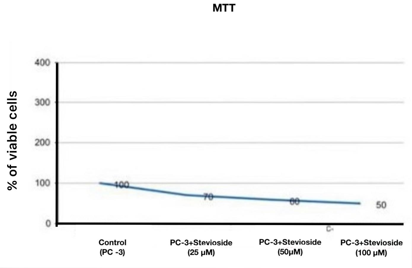
In PC-3 cells, the effect of Stevioside extracts on cell viability was analyzed through MTT assay (Figure 1) with a significant decrease in the percentage of viable cells with an increase in concentration, while the Mcl-1 gene (Figure 2) with a decrease in fold change with the rise in concentration. Caspase 3 (Figure 3) with a significant increase in fold change with an increase in concentration indicates effective apoptosis and also inhibits the Bcl-2 gene with a decrease in fold change with a rise in concentration (Figure 4). Calculated mean ± SD by one-way ANOVA test represented significance with p<0.05 which indicates Stevioside as an effective Anticancer agent.
|
|
|
Figure 1. Assessment of Cell viability |
|
|
|
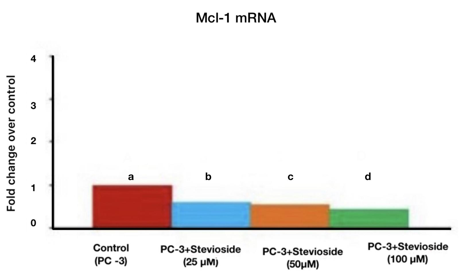
Figure 2. Graph depicts X-axis with PC-3 cell line treated with Stevioside concentration at different micromoles with a) Control (Red), b) 25μM (Blue), c) 50μM (Orange), and d) 100μM (green) and Fold change over control is indicated on the Y-axis. With respect to the expression of Mcl-1mRNA, Stevioside's anti-cancer activity significantly decreased when the concentration reached 100μM. |
|
|
|
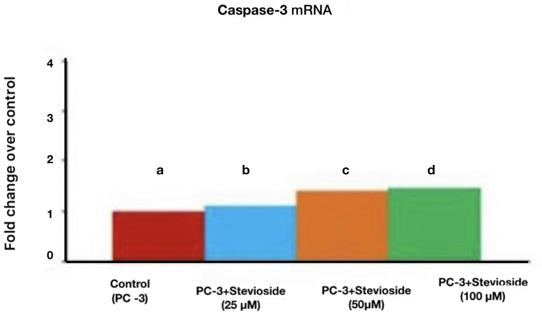
Figure 3. Graph depicts X-axis with PC-3 cell line treated with Stevioside concentration at different micromoles with a) Control (Red), b) 25μM (Blue), c) 50μM (Orange), and d)100μM (green) and Fold change over control is indicated on the Y-axis. With respect to the expression level of the Caspase 3 gene, Stevioside's anti-cancer action significantly enhanced as the concentration reached 100μM. |
|
|
|
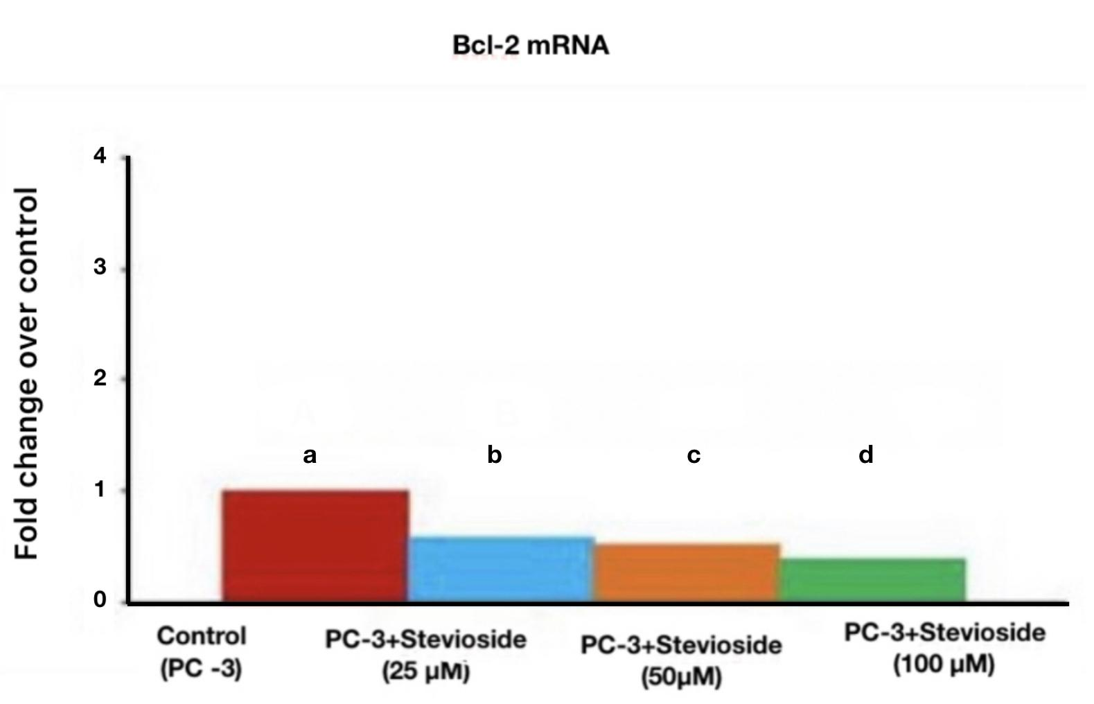
Figure 4. Graph depicts X-axis with PC-3 cell line treated with Stevioside concentration at different micromoles with Control (Red), 25μM (Blue), 50μM (Orange), and 100μM (green), and Fold change over control is indicated on the Y-axis. With respect to the expression of Bcl-2 mRNA, Stevioside's anti-cancer effect significantly decreased when the concentration reached 100μM. |
The anticancer activity of Stevioside was analyzed using a human prostate cancer cell line in vitro which had shown a positive correlation with caspase 3 and a negative correlation with Mcl-1 and Bcl-2 genes enacting its anticancer property. Earlier work in castration-resistant prostate cancer showed that chemotherapeutic drugs targeting Mcl-1 mRNA promoted cell death in response to DNA damage (CRPC).[11]
In this work, an increase in Stevioside concentration dramatically reduces Bcl-2 mRNA gene expression when compared to the control. Previous studies demonstrated that Stevioside caused apoptosis to occur.[12] Prostate cancer cells can be treated with stevioside's anticancer characteristics without suffering any negative side effects.[13, 14]
In the current investigation, the expression of the Caspase-3 mRNA gene significantly increased as the Stevioside concentration rose. Apoptosis indicators were upregulated in previous work using silver nanoparticles aided by Stevioside. Thus, demonstrating Stevioside's effectiveness against hepatocellular carcinoma as a stable prospective anti-cancer medication.[15] Prostate cancer cells are resistant to the anticancer effects of Stevioside, which is found in the Caspase-3 mRNA gene and can be treated or cured with few side effects.
The biological and pharmacological properties of Stevioside and related substances have been extensively studied. There are, however, some crucial information gaps that prohibit it from receiving the Food and Drug Administration's "Generally Recognized as Safe (GRAS)" certification.[16] Its usage in the United States is therefore restricted to that of a "dietary supplement," even though it has been approved for use as a food additive in several other nations. The anticancer effects of Stevioside in preventing prostate cancer are explored in this study. Prostate cell lines can be replaced with other cell types and the number of genes used to create Stevioside can be increased in upcoming research works.
According to the results of this investigation, the Stevioside extract considerably and strongly (by a significant fold change) suppresses the proliferation of cancer cells. The results imply that Stevioside may be turned into a natural prostate cancer medication and further investigation is necessary to establish the daily intake of Stevia products that is both safe and beneficial to the health of the human body.
The authors are thankful to Saveetha Institute of Medical and Technical Sciences (SIMATS), Saveetha Dental College and Hospitals, Saveetha University for giving a platform to conduct the study.
None.
The present project is funded by
None.
|
||||||||