Deniz Publication
Clinical Cancer Investigation Journal
ISSN Print: 2278-1668, Online: 2278-0513
ISSN Print: 2278-1668, Online: 2278-0513
Absence of Synergistic Effect of Toluidine Blue and Cytomorphometry in Discriminating Dysplasia in Oral Exfoliative Cytology
Yashi Shrivastava1, Monal Yuwanati2*, Narayanan Ganesh3
1Department of Oral Pathology and Microbiology, People’s College of Dental Science and Research Centre, People’s University, Bhopal. 2Department of Oral and Maxillofacial Pathology, Saveetha Dental College and Hospitals, Saveetha Institute of Medical and Technical Sciences, Saveetha University, Chennai, 600077, India. 3Department of Research, Jawaharlal Nehru Cancer Hospital & Research Centre, Idgah Hills, Bhopal, MP, India.
Abstract
Numerous tools and techniques such as vital stain, autofluorescence, exfoliative cytology, histomorphometry, etc. were explored in the detection of dysplasia, but have variable sensitivity and specificity. The present study evaluated the effect of toluidine blue in increasing the accuracy of histomorphometry analysis in detection of the dysplasia in OPMDs on exfoliative cytology. An experimental, observational, cross-sectional research was performed on patients attending dental hospitals and in oral health check-up camps suspected of having OPMDs. Cytology smear was collected using the standard protocol before and after the application of a toluidine blue stain over the lesion. Two pathologists performed the dysplasia grading and histomorphometry analysis. Chi-square and t-tests were carried out (p<0.05). The study observed a positive correlation between toluidine blue positivity and dysplasia in OPMDs. The toluidine blue stain sensitivity, specificity, Positive Predictive Value, and Negative predictive value were 39.53 %, 97.56, 97.14, and 43.58% respectively. Toluidine blue before the cytology smear does not improve the diagnostic accuracy of cytomorphometry. Further, there was no significant variation in cellular and nuclear morphometry parameters as compared to normal mucosa. Toluidine blue stain does not improve the efficacy of cytomorphometry in discriminating dysplasia on cytosmears. However, toluidine blue stain can assist clinicians in identifying potential areas of having a suspicion of dysplasia for scalpel biopsy.
Keywords: Cytomorphometry, Toluidine blue, Oral premalignant lesion, Leukoplakia, Erythroplakia, Dysplasia
Oral cancer is still a debilitating disease affecting millions of people in India. Oral cancer is a preventable disease through early identification of premalignant changes in the oral cavity in the early days.[1] Oral premalignant lesions have a higher risk of malignant transformation.[2] Clinically, these premalignant lesions are difficult to be labeled as dysplastic because not all lesions undergo malignant transformation. To catch the oral malignant lesion in its early stage, numerous tool and techniques were explored.[3] A few diagnostic tools for early detection of oral cancer include tolonium chloride or toluidine blue dye, Oral CDx brush biopsy kits, salivary diagnostics, and lastly optical imaging systems. In Past, toluidine blue (97.8 % Sensitivity; 100 % specificity) has been used to predict dysplastic changes in the mucosa.[4] However, despite its non-invasive, simple use, and cost-effective advantages, toluidine blue can produce a false positive result, especially in the case of inflammatory lesions.[5] The premalignant lesion, histologically, shows abnormal or altered nuclear and cell changes which exfoliative cytology can be easily identified.[6] Recent development in information technology has developed histomorphometry of oral premalignant lesion.[7] Considering its effectiveness and elimination of pathologist bias, not only it can prove effective in detecting dysplastic change at a very early stage but also it can be used as a follow-up or screening tool along with exfoliative cytology. However, these histomorphometric studies lacked comparative evaluation, as well as morphology values, were variable.[8] Even clinicians face problems in the early detection of this lesion, which hinder them from doing the provided proper treatment.[9] Further, follow-up is difficult due to the absence of any definitive detection tool.[3] Out above tools, toluidine blue stain and cytology are considered to be having better sensitivity and specificity. But still lack improvement in false negativity and false positivity.
To address this issue, Caruntu ID et al.
proposed a quantitative approach to the construction of a classifier for detecting the presence of dysplastic/cancerous cells on cytology smears.[10] The use of exfoliative cytology has been of importance only in the detection of cervical cancer whereas, in oral cancer, and precancer detection, it has been only an adjuvant because of its unreliability. Adding the toluidine blue vital staining increase its sensitivity but still, it does not eliminate the limitations of exfoliative cytology. Exfoliative cytology along with toluidine blue can have a synergistic effect in evaluating the morphology of cells. Adding quantitative histomorphometry can give a very interesting insight into this old but time test technique especially due to its low cost, non-invasive nature. Considering all the above-mentioned facts, the present study was conducted to know whether toluidine blue stain can improve the efficacy of cytomorphometry in discriminating dysplasia.
Materials and Methods
The observational, analytical, cross-sectional, single-blind study was conducted among representative samples between the age group of 15 to 35 years. Prior approval from the Institutional ethics committee was obtained. Samples were drawn from metro city and surrounding areas over 1 month. Initial screening for oral potentially malignant disorders as per WHO definition was conducted in oral health camps organized at various locations. The principal investigator performed the clinical examination under the supervision of a guide. Patients having suspected oral premalignant disorder/lesion was selected for inclusion. Required details we rerecorded in sheet. Patients were recruited continuously until the sample size was reached. Detailed information was provided to participants and informed consent was obtained. We excluded patients allergic to toluidine blue and unwilling to take part in the study. The dependent variable and Independent variable were the degrees of dysplasia, cytomorphometric values of cells and inflammatory lesion or process, and pathologist skill, respectively. The confounding factor considered was tobacco chewing/smoking habit and areca nut chewing, candidiasis.
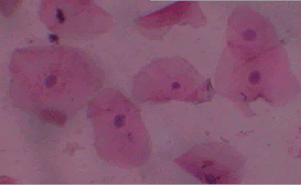
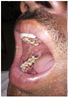
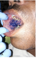
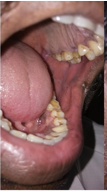
After identification of the suspected oral premalignant lesion, each participant was asked to rinse their mouth and the first cytology sample (Figure 1) was taken and labeled systematically. After that, toluidine blue staining was performed with all adequate precautions, and the finding was noted in the record sheet (Figures 2a and 2b). Another cytology smear was taken from the toluidine blue positive area and labeled. Those cases suspicious of malignant change at the time of clinical examination were also advised to undergo biopsy after the consent from the participant. Two independent pathologists were the dysplasia grading of cytology samples. After dysplasia evaluation, a cytology smear was photographed, and a re-assigned code number was to blind it for histomorphometric evaluation. Two pathologists performed the cytomorphometric analysis using morphometry analysis software (Magnus Pro). Normal mucosa for the same patient was used as positive control measurement for cytomorphometry. Data collected from the previous exercise was arranged systematically in the table and analysis was performed for the following outcome: 1. Correlation of cytology diagnosis of presence or absence of dysplasia, 2. Comparison of cytomorphometric detection of dysplasia with normal mucosa, 3. Correlation of cytomorphometry and toluidine blue staining.
|
|
|
Figure 1. Cytology smear of patients. |
|
|
|
|
|
|
a) |
b) |
||
|
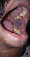
Figure 2. Toluidine blue staining a) Positive staining b) Negative staining |
|||
Results and Discussion
The study involved 126 participants with OPMDs. 99 males and 27 females were participants. The mean age was 30 yrs. Tobacco was the most common substance consumed among the participants followed by betel nuts and cigarettes. Participants with habit were having clinically identifiable oral lesions, leukoplakia (n=67), oral submucous fibrosis (n=53), erythroplakia (n=29), and Lichen planus (n=11). Cytological examination of smears from 126 participants shows the presence of dysplasia in 86 participants. These were correlated with toluidine blue staining. It was found that out of 86, only 34 were Toluidine Blue stain-positive. A descriptive analysis is given in Table 1.
Table 1. Characteristics of Study group |
||||
|
Gender |
Male |
99 |
||
|
Female |
27 |
|||
|
Habit |
Tobacco |
119 |
||
|
Betel nut |
71 |
|||
|
Cigarette |
63 |
|||
|
Duration of Habit |
yrs. |
Tobacco |
Cigarette |
Betel nut |
|
0-1 |
07 |
64 |
55 |
|
|
1-5 |
17 |
11 |
08 |
|
|
6-10 |
23 |
10 |
13 |
|
|
11-15 |
30 |
17 |
18 |
|
|
15-20 |
30 |
14 |
20 |
|
|
20-25 |
11 |
11 |
12 |
|
|
Type of lesion |
Leukoplakia |
67 |
||
|
|
Erythroplakia |
29 |
||
|
|
Oral Lichen Planus |
11 |
||
|
|
Oral Submucous Fibrosis |
53 |
||
|
Toluidine blue test |
Positive |
35 |
||
|
|
Negative |
91 |
||
|
Dysplasia |
Present |
86 |
||
|
|
Absent |
40 |
||
A chi-square test of independence was performed to examine the relationship between the presence of dysplasia and toluidine blue positivity (Table 2). The relation between these variables was significant, X2 (1, N = 126) = 18.6656, p = .000016). Dysplastic lesions were more likely to show positivity than non-dysplastic (Normal/Benign) lesions. The toluidine blue stain sensitivity, specificity, positive Predictive Value, and Negative predictive value were 39.53 %, 97.56, 97.14, and 43.58% respectively. Toluidine blue is a vital stain taken up by the cell nucleus due to its affinity to DNA and RNA. To find whether toluidine blue stain help in increasing accuracy in the detection of dysplasia on exfoliative cytology of oral lesion after TB stain application, a chi-square test was performed. The relation between these variables was significant, X2 (N = 126) = 81.457, p <.00001) (Table 3). The Kappa value was 0.785 indicating that there was moderate inter-observer agreement on the presence of dysplasia.
Table 2. Correlation of the presence of dysplasia with toluidine blue positivity |
|||||
|
Dysplasia |
|
||||
|
Present |
Absent |
Total |
P value |
||
|
Toluidine blue staining |
Positive |
34 |
1 |
35 |
0.000016 |
|
Negative |
52 |
39 |
91 |
||
|
Total |
86 |
40 |
126 |
||
Table 3. Inter-observer agreement on dysplasia (After toluidine blue application) |
|||||
|
|
First Cytology smear |
Total |
P value |
||
|
Dysplasia |
Normal |
||||
|
Second cytosmear |
Dysplasia |
35 |
12 |
47 |
0.00001 |
|
Normal |
0 |
79 |
79 |
||
|
Total |
35 |
91 |
126 |
||
Morphometric cell values were compared between normal mucosa and dysplastic mucosa cells to find out changes in the morphometric parameter. In dysplasia, nuclear parameters increase and cellular parameter decreases with an increase in nuclear-cellular area and diameter ratio. T-test showed that there is no statistical difference in the morphometric value between normal mucosa and dysplastic mucosa (Table 4). In addition to the above, the t-test was performed to compare the mean cytomorphometric value of CA, NA, NA: CA ratio, CD, ND, and ND: CD ratio found whether toluidine blue stain has any effect on morphometric parameters. The results found that there is no significant change in the mean value of morphometric parameters after the application of the toluidine blue stain on the oral mucosa (Table 4). however, there was a statistically significant difference between morphometric values of dysplastic cells measured before and after toluidine blue stain application (Table 4). CA, NA, CD, and NCD showed significant changes in values. Decrease in CA, NA, and CD whereas increase ND, NCA, and NCD was seen after the application of toluidine blue stain. This indicated that toluidine blue might not assist the cytomorphometry discriminatory efficacy in detecting dysplasia.
Table 4. Comparions of cytomorphometric parameter for normal mucosa and dysplastic mucosa before and after TB application. CA, NA, CD, and NCD showed significant changes in values |
||||||||||
|
Normal Mucosa |
Dysplasia |
|
|
|||||||
|
Before TB application |
After TB Application |
Before TB application |
After TB Application |
Normal vs Dysplasia (After TB Application) |
Pre and Post TB application (dysplastic mucosa) |
|||||
|
Mean |
SD |
Mean |
SD |
Mean |
SD |
Mean |
SD |
t-test (p-value)† |
p-value |
|
|
CA |
687.8532 |
108.3428 |
620.2732 |
81.16636 |
748.1616 |
60.16166 |
610.5762 |
87.02875 |
0.1541 |
0.009* |
|
NA |
16.0594 |
1.173549 |
14.2784 |
1.012995 |
15.5528 |
1.244192 |
14.826 |
1.641975 |
0.2632 |
0.032* |
|
NA:CA |
0.02392 |
0.004922 |
0.023456 |
0.004298 |
0.020913 |
0.002626 |
0.024679 |
0.004246 |
0.1313 |
0.334 |
|
CD |
33.794 |
1.783708 |
32.3504 |
1.970659 |
34.4218 |
3.305812 |
29.644 |
4.045592 |
0.3592 |
0.011* |
|
ND |
4.446 |
0.386546 |
4.5468 |
0.300343 |
4.2592 |
0.730135 |
4.241 |
0.713402 |
0.3133 |
0.749 |
|
ND: CD |
0.131974 |
0.014864 |
0.140967 |
0.012500 |
0.123114 |
0.012186 |
0.143854 |
0.023911 |
0.1664 |
0.018* |
† Comparison of After TB application values of normal mucosa and dysplastic mucosa, * Statistically significant; CA-Cellular Area, NA-Nuclear Area, CD-Cellular diameter, ND-Nuclear Diameter, TB-Toluidine Blue
Oral premalignant lesions have the potential to transform into malignancy. Early detection of additional measures such as stoppage of habits is key to preventing malignant changes in the lesion. Exfoliative cytology is an easy, non-invasive, patient-compliant technique used in past for the early detection of cervical and oral malignancy. However, it is subjected to variability due to various factors such as the training and experience of cytologists. Toluidine blue vital stain was used as an adjunct in assisting the clinician to identify the location showing dysplastic changes. However, it is also subjected to variability due to tissue changes or processes such as inflammation or benign proliferative activity. Considering the situation, a computerized method such as cytomorphometric analysis of cells has been utilized and found to have a role in detecting dysplasia objectively. This study was undertaken to find the efficacy of cytomorphometry along with toluidine blue vital staining in the detection of dysplasia in oral premalignant lesions at an early stage.
Cytological diagnosis of dysplasia is based on cellular and nuclear features of cells. It includes an increase in nuclear hyperchromasia, an increase in nucleus size, a decrease in cellular size, and an altered nuclear-cytoplasmic ratio. It was observed by a pathologist on a microscope to determine the cytological diagnosis of a dysplastic lesion. 35 cases were showing dysplasia on exfoliative cytology. Exfoliative cytology collects the superficial cells, which gives a slight drawback to the technique. However, sometimes can collect deeper cells depending upon the type of brush such as OralCDx. OralCDx brush is costly and it may not be feasible and affordable in rural areas. The toothbrush was used as cytobrush to check the efficacy of cytology to detect dysplasia. The authors found that brush biopsy has a good correlation with histopathological diagnosis of dysplasia and malignancy. The studies that modified the brush have reported cytology as a sensitive and reliable tool in poor resource areas for dysplasia detection. However, variation in cytological interpretation especially false negative results can be due to various factors such as sampling error, laboratory fixing, staining error, pathologist experiences, etc. The false negative rate in exfoliative cytology is very high.[7] One of the important reasons for it could be improper site selection.[11] Toluidine blue vital stain can assist in determining the suspicious site for sampling thereby helping in reducing the false negative case.[12] Parekh et al. conducted a case-control study to determine the usefulness of toluidine blue staining in selecting sampling sites for biopsy in potentially malignant disorders.[13] They reported that toluidine blue stain positivity has a good correlation with dysplasia detection and it can be an excellent tool to rule out potentially malignant lesions/disorders. In the present study, 27 % of the case were toluidine blue positive. The sensitivity; specificity, positive Predictive Value, and Negative predictive value were 39.53 %, 97.56, 97.14, and 43.58% respectively. Low sensitivity was reported in previous studies.[13-15] It could be attributed to surface keratosis or toluidine blue application for a long time.[13-15] The low sensitivity in the present study could be due to high number of leukoplakia and oral submucous fibrosis case, which shows surface keratosis.
A new approach toward oral cytology to improve exfoliative cytology was taken recently. It involved a modified brush and oral rinse technique.[16, 17] However, subjective bias on part of the pathologist is a prime factor in diagnosis especially if the pathologist lacks training and experience in cytology interpretation. Intra-observer variability is present in exfoliative cytology.[18] These studies have reported poor to moderate agreement which was similar to the present study.[19] The premalignant lesions show less inter-observer agreement. The researcher performed a computerized analysis of cytology to remove the subjective bias and improve the inter-observer agreement.[7, 20, 21] The histomorphometric analysis of exfoliative cells from normal mucosa and oral premalignant lesions in these studies was found to be effective in differentiating between dysplastic and non-dysplastic cells, thereby helping in the diagnosis of the dysplastic lesion. These reported a decrease in CA and CD; an increase in NA, and ND, and an increase NACA, and NDCD ratio in dysplastic cells as compared to normal cells. There is an increase in nuclear parameters and a decrease in cell parameters from normal to an increase in the grade of dysplasia. This correlates with different features given in various grading systems such as WHO or the Binary grading system. Although these studies reported variation in cytomorphometric values between normal dysplasia, the present study did not find any variation. This could be due large number of cases in the early stages of the cytology technique limitation to collect only superficial cells rather than deeper cells.
Toluidine blue stain was utilized as a vital stain to find its advantage in increasing the accuracy of cytology diagnosis in dysplastic lesions.[22] Ratna et al. measured morphological parameters in the smears obtained before and after the toluidine blue application.[22] They observed the maximum enhancement in cytological smears post-toluidine blue application. Among the individual parameters, nuclear pleomorphism exhibited the greatest significant difference. There was a positive relation between toluidine blue application and cytological diagnosis in the present study. A similar effect of toluidine blue stain in enhancing staining characteristics were reported by other studies.[23] However, this needs to be explored with large samples in standardized settings such as randomized case-control studies using histopathology as the gold standard.
Conclusion
The study observed a positive correlation between toluidine blue positivity and dysplasia. The present study found that dysplastic lesions were more likely to show positivity than non-dysplastic (Normal/Benign) lesions. The toluidine blue stain have low sensitivity (39.53 %) and Negative predictive value (43.58%). However, toluidine blue might not assist pathologist in detecting dysplasia on cytology smears. Simiarly, it does not aid histomorphometry in discriminating dysplasia on cytosmears.
None.
None.
This work was partially supported by the STS Program of the Indian Council of Medical Research, New Delhi, India [Reg. No: 2019-04212].
|
||||||||